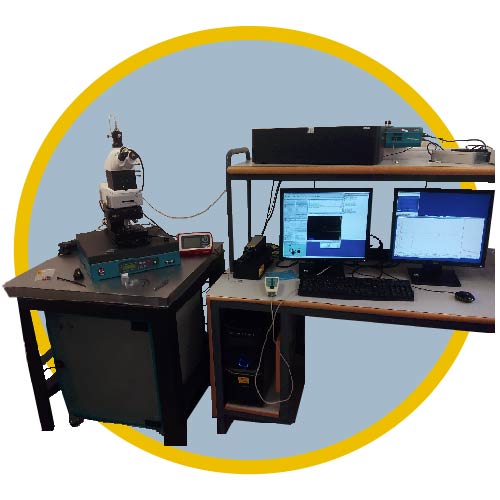
RAMAN AFM
La microscopia Raman es una técnica especializada de microscopía óptica en la que el material estudiado se ilumina con una luz monocromática (un láser) y la luz difundida por el material es analizada con un microscopio óptico confocal acoplado a un espectrómetro Raman. Una pequeña cantidad de la luz difundida experimenta un ligero cambio de frecuencia, que es característico de los enlaces químicos o de las moléculas presentes en el material. El análisis de las frecuencias de luz difundida (espectroscopía Raman) da información sobre la composición química del material y otros aspectos como estructura cristalina, orientación, grado de orden, tensión mecánica o temperatura.
RAMAN AFM EQUIPMENT
WITec combined confocal micro Raman imaging and Atomic Force Microscope (AFM) system alpha300 RA
Alpha300 RA (Witec) is an excellent combination of comprehensive surface characterization on the nanometer scale (AFM) with confocal Raman dispertion. A motorized table allows automatic mappings, line scannings or signal variations in depth. Our lasers supply different monochromatic wavelengths by means of optical fibers. The wavelength at 633 nm which offers low fluorescence on metallic substrate whilst retaining relatively high Raman intensity and 432 nm wavelength is useful for Raman resonance experiments and for the study of carbon materials
Highest sensitivity:
Gaussian Beam
High throughput configuration:
Piezo-Stage 200×200
Research grade optical microscope:
6x objective turret
Video system:
eyepiece color video camera
LED white-light source for Köhler illumination
Manual sample positioning:
x- and y-direction 25mm travel
0.25 mm pitch
Resolution:
< 1 μm
Piezo-driven scan stage:
scan range 200 x 200 x 20 μm
scan resolution: 1 nm
Position accuracy:
< 2 nm in x- and y-direction
< 0.2nm in z- direction
Lateral resolution:
diffraction limited, typically < 350 nm FWHM (532 nm excitation, 100x NA 0.9 objective, 50μm diameter pinole
Unrivaled depth resolution:
down to < 800 nm FWHM (532 nm excitation, 100x NA 0.9 objective, 25μm diameter pinhole)
Samples:
Maximal 120 mm in x- and y-direction, 25 mm in height
Laser:
Frequency doubled Nd: YAG laser, 532 nm excitation wavelength
Power:
40 mW at laser output. 633 nm excitation wavelength, 18 mW power at laser output
Spectrometer and detector:
Ultra High Throughput Spectrometer (UHTS) lens based 2 gratings: 600 and 1800 line/mm Spectral resolution <0.8 cm-1/CCD pixel Spectral cut off < 85 cm-1 Quantum efficiency of CCD detector at 532 nm better than 95%.

